Detectable changes in the brain after ILF neurofeedback: There is now imaging evidence for changes in brain connectivity.
not is a specialist magazine aimed at patients with traumatic brain injuries, stroke, and other acquired brain injuries - while simultaneously offering insights for healthcare professionals, relatives, and interested parties. The magazine provides well-founded articles, practical tips, and current developments in the field of neurorehabilitation.
In March, the magazine published an article on the topic of ILF Neurofeedback in collaboration with BEE Medic:
Making changes in the brain visible
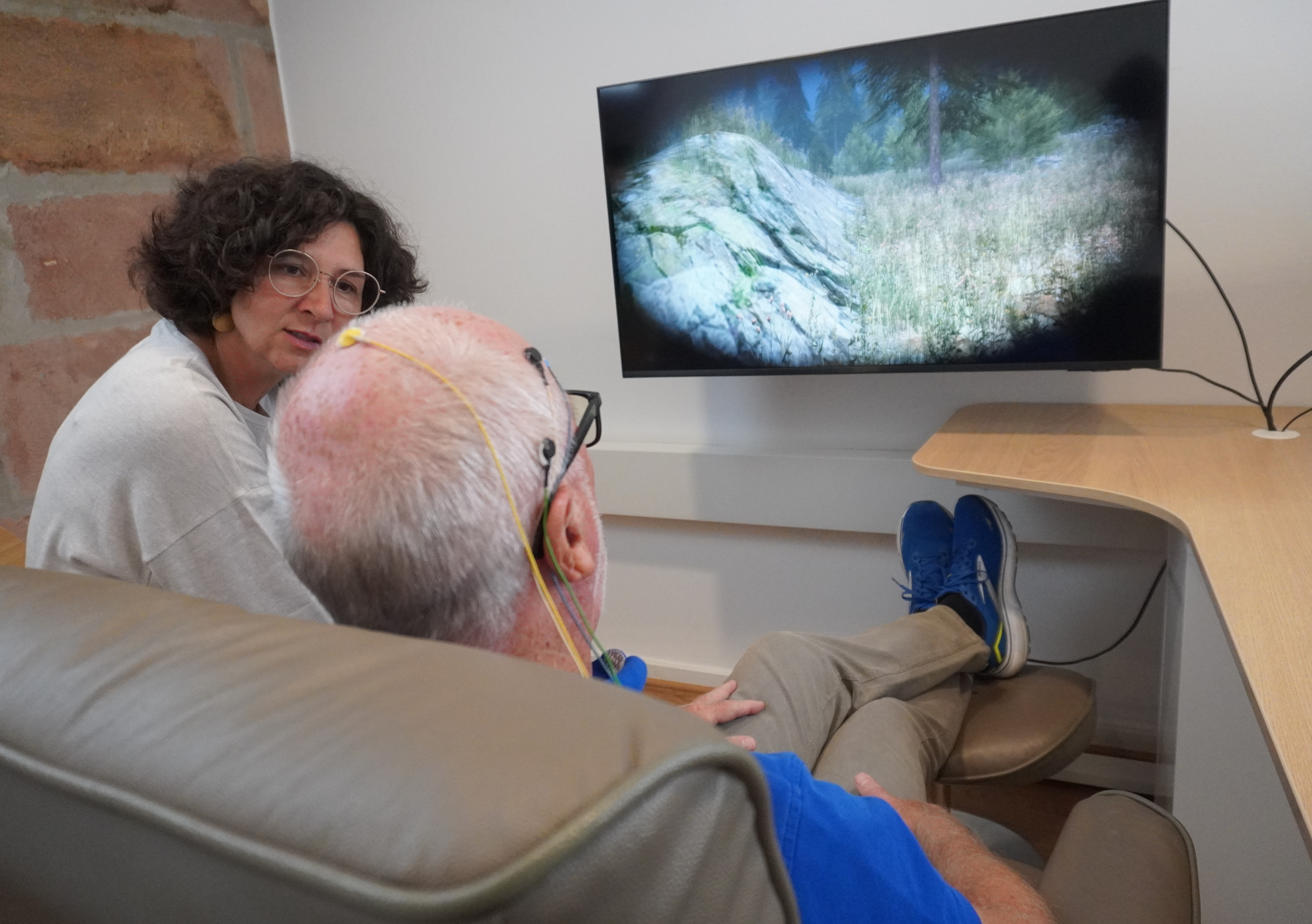
In neurological rehabilitation, neurofeedback is increasingly being used. This refers to methods in which the electrical activity of the brain is measured via electrodes on the surface of the scalp and analyzed using an electroencephalogram (EEG). The information obtained is translated into real-time feedback, for example through visual and auditory changes in a film or animation. In this way, the brain continuously receives information about its own activity. This feedback can enable changes in the organization and regulation of neuronal activity.
Symptom-based application
One possible approach is Infra-Low-Frequency Neurofeedback (ILF neurofeedback). Extremely slow changes in brain activity are included in the feedback, which are associated with higher-level processes of self-regulation and stabilization. ILF neurofeedback does not rely on conscious tasks but works predominantly on an unconscious level. In practice, it is applied based on symptoms. The starting point is specific complaints, understood as an expression of limited regulatory capacity. ILF neurofeedback is not used in isolation but combined with classical frequency band information. Only this combination constitutes the complete ILF neurofeedback protocol.
Imaging methods
In this context, imaging methods provide an additional perspective. Functional magnetic resonance imaging (fMRI) plays a central role here. It allows indirect measurement of changes in brain activity by assessing blood flow and oxygen supply in the brain. This makes it possible to map functional brain networks.
Against this background, a key question arises:
Can changes in the brain after neurofeedback be objectively demonstrated not only through symptom progression but also using imaging methods?
Extensive body of research
Numerous clinical studies already exist on the effectiveness of neurofeedback-based methods, including ILF neurofeedback. What has largely been missing so far, however, are imaging data showing whether and how different components of an established ILF neurofeedback protocol are reflected in the brain. A study published in December 2025 in the journal NeuroImage addresses exactly this question. The aim of the investigation was not to evaluate treatment outcomes but to examine whether objective changes in the brain can be detected after a single neurofeedback session and whether these depend on which signal components are used.
A total of 135 healthy adults participated in the study. The focus was therefore on fundamental neurophysiological effects rather than clinical treatment outcomes. Participants were divided into groups and received different forms of neurofeedback, using either individual signal components or their combination. Functional MRI was used before and after the session to assess changes in brain connectivity.
Results
The results show a clear pattern. Stable and statistically robust changes in functional connectivity were observed exclusively after application of the full ILF neurofeedback protocol, meaning the combination of classical frequency band information and infra-low-frequency signals. When the signal components were applied separately, no comparable stable changes could be detected.
Conclusion
Although the present study does not make statements about clinical effectiveness, its significance for understanding neurofeedback is considerable. Using imaging data, it shows that ILF neurofeedback is associated with stabilization and increased connectivity of functional brain networks.
The study does not add further effectiveness data to existing clinical studies but instead provides an imaging-based perspective on the underlying neurophysiological processes. For patients, this can serve as an important point of orientation. Neurofeedback can thus be understood not only through subjective experience but also through objectively measurable changes in the brain under clearly defined experimental conditions.
Read more about the fmri study here.